


Varicose veins during pregnancyis the ectasia of venous vessels that occur during pregnancy and are pathogenetically related to it. Violence manifests itself with paresthesia, pain in the lower extremities and external genitalia, swelling, muscle spasms, trophic skin lesions. The diagnosis is made by ultrasound angioscan. Treatment during pregnancy is usually limited to compression therapy with sleep and rest, physical activity, and improved nutrition. Perhaps the appointment of phlebotonics, phleboprotectors, anticoagulants, antiplatelet agents. Surgical treatment is usually used after childbirth.
General information
Varicose veins (varicose veins) are one of the most common vascular diseases associated with pregnancy. According to research, 15-20% of people suffer from venous pathology, 2/3 of them are women, and 60-80% of cases of venous ectasia are caused by pregnancy. The disease is generally diagnosed for the first time, and 75% of them are under 30 years old. In more than two-thirds of cases, varicose vein clinic begins after the 20th week of the first pregnancy. The urgency of timely detection of varicose veins is associated with the possibility of fetoplacental insufficiency and the risk of fatal thromboembolic complications in the absence of adequate therapy.
Reasons
Given the statistics on the frequency of varicose veins during pregnancy, most specialists in obstetrics and gynecology consider the disease as a complication of pregnancy. The predisposing factor that causes vascular ectasia in 91% of patients is a genetically determined failure of the middle vascular membrane, in which the amount of collagen is reduced and the amount of polysaccharides is increased. Facilitates the development of varicose veins in women prone to constitution during pregnancy:
- Increased blood circulation. In pregnant women, the increase in BCC ranges from 30-50% (when bringing 1 child) to 45-70% (if the child has 2 or more fetuses). This compensation mechanism allows the child, the woman's vital organs and the fetoplacental system to receive adequate blood supply.
- Hormonal regulation during pregnancy. During pregnancy, the ovaries and placenta intensively secrete progesterone and relaxin. Under the influence of these hormones, the smooth muscle fibers of the vessels relax and the structural reconstruction of connective tissue takes place. As a result, the vessel wall struggles worse with increasing intravenous pressure.
- vasoconstriction during pregnancy. The growing uterus squeezes the inferior vena cava and iliac veins. Blood flow from the pelvis and lower extremities is disrupted, intravascular pressure increases, and the venous walls become elongated. The effect of this factor plays a key role in the formation of varicose veins after the 25th week of pregnancy.
- Changes in the hemostasis system. As labor approaches, the fibrinolytic activity of the blood decreases and the number of coagulation factors increases. This adaptation mechanism aims to reduce the amount of physiological blood loss during labor. This increases the likelihood of thrombosis of pathologically altered vessels.
An additional etiofactor that contributes to the onset of varicose veins in pregnant women is a decrease in physical activity. Blood stasis in the legs and pelvis increases with insufficient skeletal muscle function. The condition is aggravated when the patient has excess body weight, which is a further increase in the volume of blood circulating in the vascular bed.
Pathogenesis
The starting point in the development of varicose veins during pregnancy is a violation of the compensatory capacity of the valve apparatus of the venous network. Because BCC is a mechanical barrier to growth and exit from the lower extremities, when the main arteries constrict, the blood shows increased pressure on the vessel wall. Genetically inherited connective tissue fiber deficiency increases with the relaxation of vascular smooth muscle under the influence of progesterone. As a result, the vascular lumen dilates, the valves stop closing, and blood collects in the vascular system of the lower extremities. As the disease progresses, the pathological process can spread to the vulvar ring, vagina and small pelvic vessels.
Classification
The main criteria for the systematization of varicose veins are the anatomical distribution of venous stasis and the severity of the disease. This approach allows different treatment regimens to be chosen for different variants of the disorder. Taking into account the involvement of different organs in the process, varicose veins of the lower extremities, vulvar varicose veins, varicose veins of the pelvic organs are distinguished. Depending on the severity of clinical symptoms, the following stages of dilation of the venous vessels of the lower extremities are distinguished:
- Compensated varicose veins. There are no external signs of vascular ectasia, and the pregnant woman reports leg fatigue by the end of the day, and discomfort in the calf muscles during exercise and brisk walking.
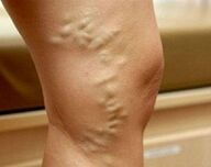
- Subcompensated varicose veins. Vein patterns ("stars") appear on the skin. In the evening the legs swell, at night there are cramps, numbness, pain. Bruises and scratches take longer to heal than usual.
- Decompensated varicose veins. The patient is constantly worried about pain in the legs, swelling increases. The veins are noticeably enlarged and nodular. The skin is hyperpigmented. There are signs of eczema and trophic diseases.
In pregnant women, the disease with pelvic varicose veins also develops in stages. In the first stage, the diameter of the affected vessels in any venous plexus of the pelvis is not more than 5. 0 mm. With the second, the uterus or ovaries are involved in the process, the lumen of the vessels 6. 0-10. It is 0 mm. Third, it is characterized by ectasia of vessels larger than 10 mm with the total presence of all pelvic venous plexuses.
Symptoms of varicose veins
In 80-82% of patients, the disease begins with a feeling of "buzzing" in the legs, which increases during weight, tension, evening and physical exertion. The symptomatology of varicose veins gradually increases. As the disease progresses in some areas of the muscles, the pain first develops with prolonged standing and physical activity. In the most severe cases, the pain is persistent and can be so intense that a pregnant woman has difficulty moving independently. Up to 60% of patients report calf muscle loss, 40-50% loss of sensation, numbness of the legs, and up to 30% itching.
In the subcompensated stage of varicose veins, there are signs of external dilation of the superficial veins. Initially, areas of reticular vessels and telangiectasia ("mesh" and "stars") form on the skin. Later the venous pattern differs. The veins appear dilated, twisted, and eventually nodular. The spread of ectasia into deep veins is evidenced by the formation of edema in the ankle joints and lower legs. With decompensation of varicose veins, the skin of the legs appears hyperpigmented, eczema develops. If the pathology occurs long before pregnancy, dystrophy of subcutaneous adipose tissue, trophic ulcers are possible.
In 4% of patients, the disease affects the vulva, vagina and small pelvic vessels. With vulvar and vaginal varicose veins, discomfort, distance, heaviness, itching are observed in the external genitalia. There may be swelling of the perineum and labia, bleeding from the vagina after sexual intercourse. Pelvic obstruction syndrome manifests itself with pulling or pain that spreads to the lower back, sacrum, groin, and external genitalia. Dyspareunia (pain during sex) is characteristic. In severe cases, dysuria is detected.
Complications
In the absence of adequate treatment, varicose veins in pregnant women can be complicated by trophic ulcers, erysipelas, thrombophlebitis, superficial and deep vein thrombosis, pulmonary embolism during childbirth, and other large vessels. In 40-45% of cases, placental insufficiency occurs with acute and chronic fetal hypoxia. Occupational anomalies are observed in 25% of patients (weakness of the workforce, violation of the contractile function of the myometrium). With vaginal varicose veins, a massive traumatic walk is possible in the postpartum period. Nearly one-third of women at birth have placental abruption. The long-term consequences of varicose veins during pregnancy are hemorrhoids, chronic venous insufficiency and pelvic pain.Diagnostics
Diagnosis of varicose veins during pregnancy with the appearance of specific skin symptoms is generally not difficult. The tasks of the diagnostic stage are to determine the stage and localization of venous ectasia, to exclude other causes that can lead to stagnation in the vessels of the lower extremities. The most informative query methods are:
- Seat Inspection. The study shows characteristic changes in the venous vessels in the vulva and the inner thighs - ectasia, bowel, nodules. Swelling of the labia and perineum is possible. When looking in the mirror, the vaginal mucosa looks hypertrophic, cyanotic. Vaginal vaults are corrected by bimanual palpation, often painful.
- USDG of the venous system. Ultrasound examination assesses the shape and diameter, length, anatomical condition and wall condition of the vessels. The method allows to determine the zones of branching, the consistency of the valve apparatus, the openness of the vessels, the presence and direction of return. You can scan both the veins of the lower extremities and the inferior vena cava (IVC ultrasound).
- Duplex scanning of leg veins. The advantage of the non-invasive method, which combines traditional ultrasound and Doppler studies, is not only to obtain detailed information about blood flow parameters, but also to visualize the venous network. Duplex angioanalysis is used to comprehensively assess the condition of superficial, perforated and deep vessels.
Radiodiagnostic methods (varicography, selective ovarigography, increased phlebography of the extremities, pelvic phlebography, CT venography, phleboscintigraphy, etc. ) are used to a limited extent due to adverse effects on the fetus during pregnancy. In difficult cases, diagnostic laparoscopy with suspicion of pelvic varicose veins is performed with caution. Differential diagnosis of varicose veins of the legs is made by drip in pregnant women, heart failure, lymphedema, acute thrombosis of the venous system. Varicose veins of the small pelvis should be distinguished from genital endometriosis, chronic inflammatory pathology of the pelvic organs, submucosal and subserous uterine fibroids, cysts and other ovarian tumors. In addition to the observation of a midwife-gynecologist, the patient is advised to consult a phlebologist, cardiologist and oncologist.
Treatment of varicose veins during pregnancy
The main goal of therapy for varicose veins in pregnant women is to stop the development of the disorder, reduce the severity of the clinical picture and prevent possible thromboembolic complications. In addition to pharmacotherapy in the safe stages of pregnancy, non-pharmacological methods are preferred:
- Compression therapy. A woman with a confirmed diagnosis of varicose veins is advised to wear it daily during pregnancy, use elastic bandages, special compression tights or compression class 1-2 socks during childbirth and the postpartum period. Compression therapy by mechanically reducing the diameter of the superficial vessels accelerates blood flow, reduces swelling and blockage.
- Herbal phlebotonics and phleboprotectors. The effect of the use of drugs of this group is associated with an increase in venous wall tone, decreased permeability, improved microcirculation, rheological properties of blood and lymph flow. The advantage of most bioflavonoids is that they can be used during pregnancy and lactation. Phlebotonic drugs are prescribed both in tablet form and externally.
- Anticoagulants and antiplatelets. Drugs with antithrombotic activity should be used with caution in the presence of signs of increased coagulation and risk of developing DIC. Drugs that inhibit platelet aggregation and have an angioprotective effect are indicated to improve blood rheology and vascular microcirculation.
Pregnant women with varicose veins are recommended special physiotherapy exercise complexes, lymphatic drainage massage, dosed walking, daily increasing contrast shower. Dietary adjustment involves the use of foods rich in fiber and vegetable oils. Injection sclerotherapy, miniphlebectomy, crossectomy, endovasal laser coagulation and other surgical treatments are used in exceptional cases with severe forms of the disease, severe pain syndrome and complications. Often, surgery is performed at the end of the lactation period.
Delivery Tactics
The preferred method of delivery for varicose veins is natural childbirth, in which elastic bandages or compression garments are applied to a woman's lower extremities during childbirth. Patients with vulvar-vaginal varicose veins require special attention to the continuous cycle by performing a protective perineotomy. When ectasized vessels are ruptured, the damaged vessels are carefully closed with a recurrent suture of the nodal conglomerate. Caesarean section is recommended for patients at high risk of thromboembolic complications and severe vulvar varicose veins.
Forecasting and prevention
Prognosis is favorable with timely detection and appropriate therapy. As a preventative measure, it is recommended to get enough sleep and periodic rest during the day in a sleeping position, placing the feet at a 30 ° angle to a firm surface. Pregnant women with a hereditary heredity should refrain from wearing shoes with heels larger than 5 cm, limit the amount of time they sit or stand, and monitor their weight gain.
To prevent varicose veins, daily walking, reducing salt intake, taking vitamin supplements that strengthen the vascular wall are effective. Patients with varicose veins who are planning a pregnancy are more likely to undergo surgery to correct the disease.